

For most of medical history, the clitoris did not exist on paper. Anatomy books described the penis in detail — its nerves, blood supply, and inner parts. The clitoris got a sentence or two, if anything at all.
Helen O’Connell, an Australian urologist, noticed this gap early in her career and spent years filling it. Her work changed what medicine knows about the female body — and what yoni massage practitioners understand about the anatomy beneath their hands.
A Surgeon Who Noticed the Gap
O’Connell became the first female urologist in Australia in 1993. Early in her career, she noticed something odd. Preserving sexual function was standard care for male patients. For women, it was rarely considered at all.
Later, she described a textbook she used in the 1980s. It had a full chapter on the penis — its nerves and blood supply. The clitoris was not in it at all.
That absence became her drive. If no guide existed on the nerves and blood supply to the clitoris, she would create one. Her career became an act of making the invisible visible.
The 1998 Paper That Started Everything
In 1998, O’Connell published her first major study on the clitoris. Using careful dissection, she showed that the clitoris was far larger than any standard anatomy book depicted.
The external part — the glans — was only the tip. Below the surface lay a wide body of tissue that reached deep into the pelvis.
This finding challenged the maps used in medical schools for many years. The paper drew wide attention and marked the start of a long effort to redraw what was known about the female body.
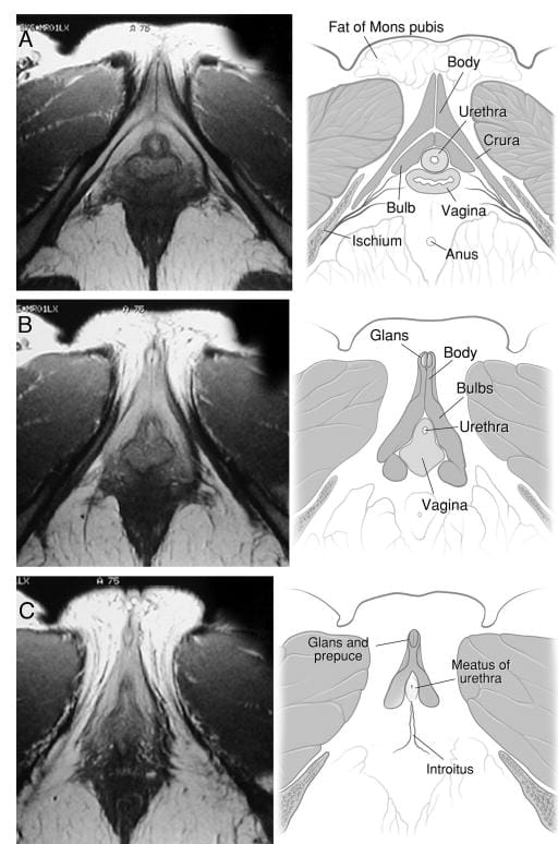
What MRI Revealed About the Clitoris
O’Connell did not stop at dissection. She wanted to see the living clitoris in a living body.
In 2005, she published a key paper in the Journal of Urology using MRI to map the clitoris in living women. MRI let her see what dissection alone could not show. The full inner size of the organ became clear. Its links to nearby parts — the urethra and the vaginal walls — could now be mapped with real accuracy.
This gave medicine its first true picture of the complete clitoris in a living body. The structure had always been there. O’Connell gave science the tools to finally see it.
auajournals.org
How Much Bigger the Clitoris Really Is
One of the clearest findings from O’Connell’s MRI work was about the tissue on either side of the vaginal opening.
For a long time, anatomy treated these as separate parts with no link to the clitoris. Her scans showed otherwise. This tissue is part of the clitoris. It fills with blood during arousal and is part of the same system as the visible tip.
Including it made the organ much larger than anyone had thought. It also helped explain why vaginal touch so often involves the clitoral system — because the clitoris is already there.
The Clitoris, Urethra, and Vagina — One System
The most far-reaching part of O’Connell’s 2005 paper was a new view of three structures: the clitoris, the urethra, and the vaginal walls. These are not separate organs with separate jobs.
All three form one linked system — sharing blood supply, nerve paths, and arousal response. This is why touching the front vaginal wall produces such a wide response. The whole CUV complex responds together.
O’Connell’s scans gave this picture a solid base for the first time. It changed how scientists and practitioners think about female arousal at its root.
MRI in the Living Body — Seeing the Clitoris in Full
O’Connell’s 2005 paper used MRI in living women to show the clitoris in its true position — something dissection alone cannot do. Scans reveal the organ as it actually sits in the body, not rotated for a dissection table.
The images showed the clitoris folding back on itself in a shape one paper called boomerang-like. That shape had never been visible before. They also showed its full links to the urethra and vaginal walls. What textbooks had shown as a flat, simple structure was now visible as a complex, three-dimensional body. The difference was stark. Later research built directly on this base.
A 2022 study from Oregon Health & Science University used microscopy to confirm over 10,000 nerve endings spread across the pelvic area — far higher than earlier estimates, and a direct sign of what O’Connell’s anatomical work had pointed to all along.
Photograph: Alana Holmberg/The Guardian
Why Medical Textbooks Got It Wrong for So Long
The gap in knowledge about the clitoris was not random. O’Connell noted something striking. The clitoris had actually grown smaller in anatomy drawings over the twentieth century. It shrank on the page across book editions while the penis grew more detailed.
This was not a neutral mistake. It showed a pattern in which female sexual anatomy was seen as less worth studying.
O’Connell’s career challenged this directly. These papers forced anatomy to correct itself. They also asked why such a large structure had been ignored for so long. That question still matters today — and the full history behind it goes back much further than the 20th century.
Her Legacy in Female Pelvic Science
O’Connell’s work created a base that later researchers built on. The nerve maps she produced guided surgeons working to protect female sexual function during pelvic surgery.
Her description of the CUV complex shaped how scientists think about female orgasm and arousal. Each researcher who came after her — studying nerve counts, fluid release, or how arousal works — built on a map she helped to draw.
Her place among the pioneers who brought real scientific care to the female body is clear and lasting.
What Her Work Means for Yoni Massage Today
O’Connell’s findings are not abstract facts. They are directly useful to every yoni massage session. Knowing that the clitoris reaches deep into the pelvis changes how a practitioner reads internal touch. Its tissue wraps around the urethra and presses against the vaginal walls.
Pressure on the front vaginal wall does not bypass the clitoris. It reaches it from the inside. Touching the sides of the vaginal opening is clitoral touch. The whole pelvic space is part of one responsive system. This is not a theory. It is anatomy — mapped, imaged, and confirmed.
Practitioners who want to work with this anatomy in a skilled way can find a full somatic framework inside the online yoni massage course.