

Understanding the internal structure of the clitoris is essential for safe and informed yoni massage. For decades, most people believed that the small visible tip was the entire organ.
Modern research has changed this completely. The clitoris is a large, complex structure that extends deep into the pelvic floor.
Studying a detailed clitoral diagram reveals a three-dimensional system — the true foundation of pelvic sensitivity and somatic practice.
The Australian MRI Breakthrough
The modern understanding of clitoral anatomy changed in 1998. Australian gynecologist Helen O’Connell used MRI scans to study the organ in living women for the first time. Her team proved that most of the clitoral tissue is internal — not external as previously believed.
By mapping the clitoris alongside surrounding pelvic organs, they produced the first accurate picture of a large erectile system capable of deep sensory response.
Before this work, anatomy textbooks had relied on outdated cadaver studies that consistently underrepresented the organ’s true size and complexity. O’Connell’s research corrected a gap that had persisted in medical education for over a century — a gap whose roots go back to Vesalius and the 16th century.

Mapping the Internal Components
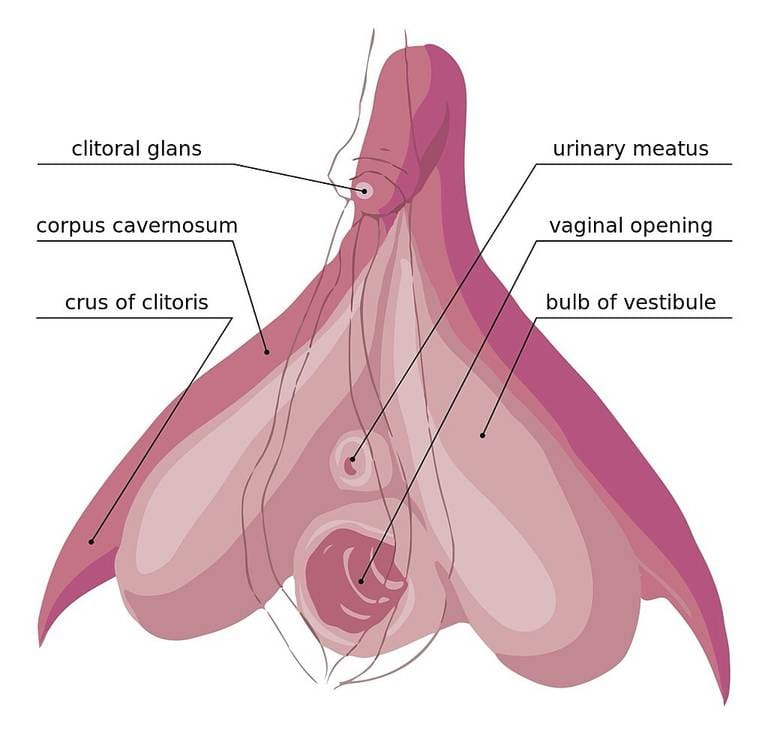
The clitoris has several parts that work as one unit. These include the glans (the visible tip), the corpus cavernosum (the erectile bodies), the crura (the legs), and the vestibular bulbs. The glans is protected by the clitoral hood — a soft, movable fold of skin that controls sensitivity.
Although the glans has the highest nerve density, it is only the tip of a much larger structure. The full length of the clitoris ranges from 8 to 12 centimeters, and in some women up to 20 centimeters.
All these parts are made of the same erectile tissue and respond as a single integrated system when arousal begins.
Homology and Embryonic Origins
The clitoris and the penis develop from the same tissue in the embryo. Both contain erectile tissue that fills with blood during arousal.
One key difference: unlike the male organ, the clitoris does not include the urethra. In the female body, the urethra sits just below, nestled between the clitoral bulbs. This creates a separate path for sensation and for biological function.
Understanding this shared origin helps explain why the clitoris has such a high capacity for arousal — it is not a lesser version of the male organ but a fully developed structure in its own right.
The Clitorourethrovaginal (CUV) Complex
Researchers Pierre Foldes and Odile Buisson introduced the term Clitorourethrovaginal complex, or CUV complex. This describes the close connection between the clitoris, the urethra, and the front wall of the vagina. These structures are so tightly linked that touching one affects the others.
In yoni massage, this area is treated as one sensory field. Tension in the vaginal wall can directly reduce the erectile response of the clitoral system. This is why external relaxation work — on the labia, the perineum, and the inner thighs — often produces changes deep inside the body that no amount of direct internal pressure could achieve alone.
Exploring the Clitoral Legs (Crura)
The crura — the legs of the clitoris — run deep inside the body along the pelvic bones. They are mostly out of reach for direct touch. Even so, they play a key role in the feeling of deep pelvic fullness. A small access point exists about one centimeter below and to the side of the glans.
In practice, the crura are influenced mainly through indirect work — massaging the surrounding tissue and bulbs to draw blood flow into the full structure.
When the entire system is engaged, the legs contribute to a broader sense of internal opening that is distinct from the sharper, more localized response of the glans.
The Vestibular Bulbs and Arousal
The vestibular bulbs sit just below the labia minora. Unlike the legs, they are reachable through massage. In many women, these tissues are dormant — numb or unresponsive due to chronic tension or lack of specific touch.
Through regular yoni massage, the bulbs can be awakened. When they fill with blood, a clitoral orgasm becomes possible without any direct touch of the external glans.
The entire internal system activates instead. This is one of the most important insights in somatic bodywork: the visible tip of the clitoris is not the only — or even the primary — pathway to deep pleasure.
Correcting Common Sexological Myths
A widespread myth in popular sexology claims that the legs of the clitoris wrap around the vagina and drive pleasure through movement. Anatomically, it is the vestibular bulbs — not the legs — that flank both the vaginal opening and the urethra.
Using an accurate clitoral diagram helps practitioners apply technique in the right areas.
The goal is to help the bulbs engorge, which creates a soft internal pressure against the vaginal walls and produces an integrated full-body response. Misinformation about anatomy leads to technique that targets the wrong structures — and misses the real potential of the organ.
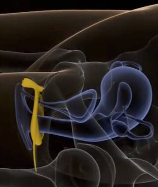
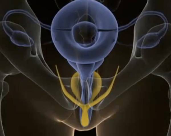
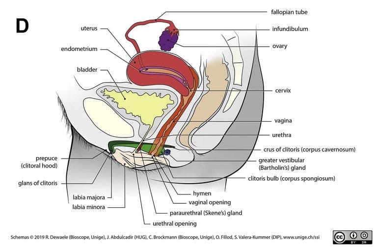
In the figures below, the clitoris with legs deep in the body and bulbs covering the urethra and vagina is highlighted in yellow. Between the bulbs is the urethra, which ends in the bladder. Below that is the vagina, into the far end of which the cervix enters.
Visualizing the 3D Anatomy
To navigate the pelvic area with confidence, a three-dimensional picture is needed.
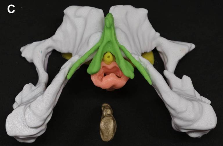
The clitoris sits like an arch over the pelvic structures. Its legs run along the bones, while the bulbs wrap around the urethra and vagina below. Seen from the side, the cervix enters the far end of the vaginal canal, positioned below the bladder.
This spatial map allows the practitioner to apply pressure that is precise, informed, and safe for the underlying organs. Diagrams and 3D models are more useful here than words alone — spending time studying them before practice is time well spent.
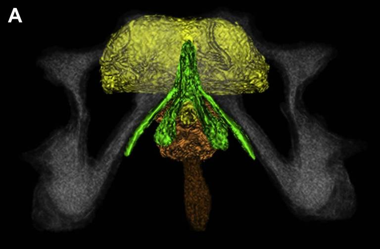
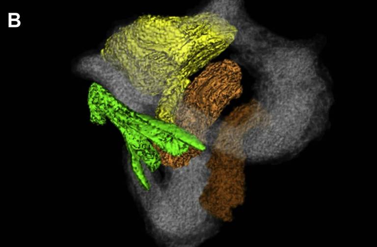
In the pictures below, the clitoris is shown in green. Its legs run along the pelvic bones.
Somatic Integration of Anatomical Knowledge
Knowing the names and locations of these structures is the first step. The deeper goal is somatic integration — letting this knowledge change how the body is experienced. When a woman learns that her clitoris is a large internal organ, her relationship with her own body often shifts.
Shame or confusion gives way to curiosity. This clarity shared between giver and receiver turns every movement in a session into an act of discovery, aimed at full pelvic presence and free energetic flow. The body learns not just through touch but through knowledge of what is being touched.
To explore how this anatomy is applied in practice, visit our yoni massage course for practitioners and partners.