

For over three hundred years, medicine ignored a small but vital organ. The tissue around the female urethra was dismissed as a useless remnant. Most textbooks did not give it a proper name.
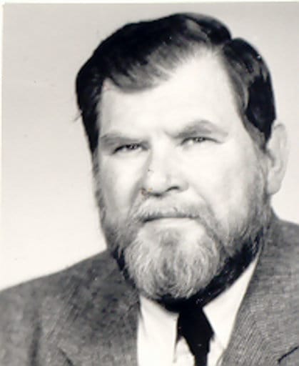
Then, in the 1980s, a forensic pathologist in Bratislava began a careful study. His name was Milan Zaviacic. Over two decades, he used electron microscopes and lab analysis to prove that the female prostate is a fully working organ — not a vestige, not a myth, and not a source of shame.
A Disputed Organ
The story of the female prostate is also the story of a mistake that lasted a century.
In 1672, the Dutch anatomist Regnier de Graaf gave the first description of glands around the female urethra. He called them a prostate and described their role in producing fluid that increases female desire.
Then, in the 1880s, Scottish gynecologist Alexander Skene published his own account. He saw only two small duct openings near the urethral entrance, naming them Skene’s glands. This narrower view stuck.
The idea that women had only two small, unimportant ducts became the standard model for a hundred years. Any tissue beyond those two openings was labeled useless. The debate was considered closed.
Milan Zaviacic and the Comenius Medical School
Milan Zaviacic began his research in 1980. He was a forensic pathologist at the Comenius Medical School in Bratislava, Slovakia. His field demands precision. Every finding must be clear, observable, and repeatable.
He brought this approach to a question most colleagues considered settled. Weekends at work, time with family sacrificed — over twenty-five years, he built a body of evidence hard to argue with.
In a 2006 radio interview, he said hard work matters more than money. Students at the faculty agreed. Soon, the female prostate became their favourite exam topic.
The Microscope Changes Everything
Zaviacic’s first step was placing female prostatic tissue under an electron microscope. What he found directly contradicted the old model. The tissue showed mature secretory and basal cells. These are the same cell types found in the adult male prostate.
Critics had claimed that female glandular cells stayed immature throughout a woman’s life.
His microscope images proved them wrong. The cells were active and equipped with the structure of a working secretory organ. They contained secretory granules and Golgi complexes. These are signs of a gland that makes and releases fluid.
This was not a dormant structure. It was a living one.
PSA as the Decisive Marker
The most powerful evidence Zaviacic produced was from lab analysis. Prostate-specific antigen, known as PSA, is the primary clinical marker for prostate tissue in men. It is used worldwide to detect prostate disease.
Zaviacic analyzed female prostatic tissue and found PSA in its secretory cells. This was not a trace finding. PSA was clearly present in the same layer as in the male prostate. Same antigen, same location, same function.
This gave his argument a clinical anchor that was hard to dismiss. The female prostate was not just similar in structure — it was the same type of organ.
https://zona.fmed.uniba.sk/
Six Types of Female Prostate
One of Zaviacic’s most useful contributions was his mapping of six types of the female prostate. He showed that the gland does not look the same in every woman.
In the most common form, most tissue sits near the external urethral opening. Around ten percent of women have a form where the tissue sits closer to the bladder neck — the area known as the G-spot. Some women have tissue spread along the full length of the urethra. Others have a small, scattered form found in about eight percent of cases. Two rarer forms show different patterns of glandular clusters.
About ninety percent of women have mature prostatic tissue capable of active secretory work.
Two Functions of the Female Prostate
Zaviacic identified two main functions of the organ.
- The first is exocrine — the gland produces PSA, fructose, and several enzymes. These compounds appear in female ejaculate. They confirm that the fluid is not displaced urine but a glandular fluid with its own chemical profile.
- The second function is neuroendocrine — the female prostate produces serotonin. Serotonin is a brain chemical involved in mood, arousal, and emotional states. This role suggests the gland plays a part in sexual arousal, not just in fluid release.
Each of these functions is real and each one matters.
The Sexological Implications
Zaviacic was direct about what his findings meant for female sexuality. In his own words, the female prostate tissue is a new erogenous zone for women.
It takes part in female ejaculation, in which the female prostate is stimulated indirectly. This indirect stimulation happens through the front wall of the vagina — the same wall where G-spot sensitivity is found.
His work links the G-spot directly to the activity of the female prostate. The fluid that many women release during deep internal touch is not urine. It is prostatic fluid.
The 2001 Recognition by FICAT
In 2001, the Federative International Committee on Anatomical Terminology met in Orlando, Florida.
Based on Zaviacic’s work, the committee voted to include the term female prostate in international terminology. The use of Skene’s glands and para-urethral ducts was officially prohibited in formal scientific publications.
In October 2008, the updated edition of Histological Terminology was published with the term female prostate included. After more than three hundred years of debate, the organ had a name. The work was done.
What This Means for Somatic Practice
Understanding the female prostate changes how we approach yoni massage. G-spot work is not stimulating a vague spot on the vaginal wall. It is applying indirect pressure to a real glandular organ with known structure, known chemistry, and known functions.
The variation between women — why some produce visible ejaculate easily and others do not — is explained by Zaviacic’s six types of the gland. In practice, most yoni massage sessions produce visible signs of this organ at work.
After deep internal touch of the front vaginal wall, the milky white fluid of female ejaculate is often visible on the practitioner’s disposable glove — a direct sign that the female prostate has been reached and activated.
To learn how to work with this anatomy step by step, explore our complete yoni massage program.